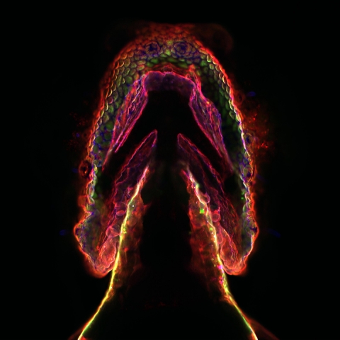
Cell surfaces are decorated with complex carbohydrate structures termed glycans, which appear as modification of lipids and proteins that reside within the plasma membrane. The ensemble of cell-surface glycans changes as cells undergo normal physiological changes such as development as well as malignant transformations in many cancers, and the ability to image these changes in glycosylation could shed light on the roles that glycans play in these important processes. The Bertozzi lab has developed a two-step chemical strategy that enables the imaging of these glycans within living systems, involving (1) the metabolic labeling of the glycans with unnatural sugars bearing a unique chemical group, the azide, and (2) the selective targeting of azide residues within cell-surface glycans with so-called "bioorthogonal" probes bearing imaging agents, including phosphine and cyclooctyne reagents. Shown is a ventral view of a 3-day-old zebrafish embryo that has been labeled using this strategy with many different color reagents, with each color representing a snapshot of the ensemble of mucin glycans at a particular point in the organism's development.