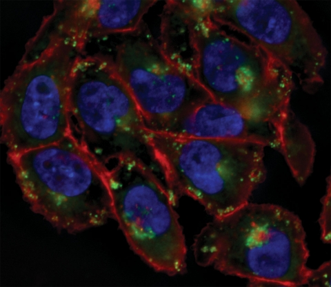
Cell surfaces are decorated with complex carbohydrate structures termed glycans, which appear as modification of lipids and proteins that reside within the plasma membrane. These glycans undergo constant remodeling, yet unlike their protein counterparts, direct imaging of glycan dynamics within living systems has not been possible. The Bertozzi lab has developed a two-step chemical strategy that enables the imaging of these glycans within living systems, involving (1) the metabolic labeling of the glycans with unnatural sugars bearing a unique chemical group, the azide, and (2) the selective targeting of azide residues within cell-surface glycans with so-called "bioorthogonal" probes bearing imaging agents, including phosphine and cyclooctyne reagents. Shown is a population of Chinese hamster ovary cells that have been labeled using this strategy. Colored in red are the glycans, and counterstains for the nucleus (blue) and Golgi apparatus (green) are also shown.